Our merge with PRP beings the ease of being able to book directly with your location of choice online.
Once you have been redirected to PRP (by clicking below), please click on Book Appointment or Book Online.

Our merge with PRP beings the ease of being able to book directly with your location of choice online.
Once you have been redirected to PRP (by clicking below), please click on Book Appointment or Book Online.
What is it?
MRI stands for Magnetic Resonance Imaging. It uses a large magnet to provide detailed images of your internal organs/anatomy. 
Why have it?
The reason for the test is best explained to you by your referring doctor. It can be used to diagnose numerous conditions.
How long does it take?
About 20-45 minutes (depending on the region being scanned).
What do I have to do?
There is no preparation for an MRI, however you must ensure there is no metal on your person, this includes jewellery and clothes. A gown can be provided if necessary. You must inform the receptionist or radiographer if there is the possibility of any foreign bodies within your anatomy, e.g. shrapnel from accident, fragments from metal work etc.
Where is it performed?
3T MRI is located at 105 Main Street (entrance at rear of building).
If you require any further information, or have any questions, please ask the receptionist, radiographer or radiologist before the examination is performed.
Alternatively, you can contact the practice on 9622 2292 or via email.
What is it?
A biopsy is a procedure in which samples of tissue are obtained via a needle. It is done with the help of imaging equipment so that the needle can be inserted directly into the location of the abnormality.
Why have it?
The procedure will allow accurate diagnosis of the problem, usually a lump. It will hopefully confirm the nature of the problem.
How long does it take?
It depends on what area is being examined – between about 20-30 minutes. The specimen is sent to a pathology lab which can take up to three working days to analyse the specimen and return a result. You can contact your referring doctor regarding these results.
What do I have to do?
There is usually no preparation but it is best not to have just eaten.
You will be required to sign a consent form as it is an invasive procedure.
What does the procedure involve?
The procedure may be done under either CT or ultrasound guidance depending on which is appropriate for your problem.
In nearly all cases, local anaesthetic will be given to numb the area. Unfortunately the anaesthetic may sting a little. Once this is done a further needle, or needles, will be inserted into the lump to obtain the cells necessary for diagnosis – this may be done a number of times.
Afterwards you may need pain killers e.g. Panadol, as there may be some pain. Also, a small bruise may appear. It is best not to take Aspirin as this may increase bruising.
Areas commonly biopsied (all performed at Western Imaging Group) include thyroid, breast, lymph node and abdomen.
Where is it performed?
All biopsies are performed at our Campbell Street rooms.
If you require any further information, or have any questions, please ask the receptionist, radiographer or radiologist before the examination is performed.
Alternatively, you can contact the practice on 9622 2292 or via email.
What is it?
CT (computed tomography) is a means of looking inside your body using x-rays, in cross section i.e. like slices of bread in a loaf. You will simply lie still on a bed and part of you will move into a large doughnut like 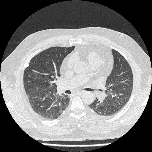 structure.
To become claustrophobic is extremely uncommon.
Why have it?
The procedure will often allow accurate diagnosis of the problem. CT however does not have all the answers and some diseases may not be able to be detected. Other imaging tests, e.g. ultrasound, may be necessary for further diagnosis.
How long does it take?
The actual scan time is usually only seconds. The computer then must reconstruct the images and then the films must be printed.
What do I have to do?
For certain tests, you will be required to sign a consent form and answer some questions, for example, regarding your allergies (if you have any) and medications.
For some scans it will be necessary for you to fast for a short period of time. For others, usually abdominal scans, it will be necessary for you to drink some special CT contrast about 1 hour prior to the examination. For some scans, an injection of contrast medium may need to be given and this will provide additional diagnostic information.
Are there any side-effects?
You are exposed to a small dose of radiation which is extremely unlikely to cause side-effects.
If you have an injection, you may get a warm feeling, bad taste in your mouth or urge to go to the toilet. These are all temporary, lasting only seconds. Nausea may occur. Itchiness or rash or more serious reactions are not common. If you are pregnant, or think you might be, please inform the radiographer before the test.
Where is it performed?
CT scans are performed at our Campbell Street, Marsden Park and Plumpton rooms. All CT guided spinal pain therapy is performed at Campbell Street.
If you require any further information, or have any questions, please ask the receptionist, radiographer or radiologist before the examination is performed.
Alternatively, you can contact the practice on 9622 2292 or via email.
What is it?
Dental imaging includes OPG (orthopantogram) and cephalometry. These x-rays image the teeth and 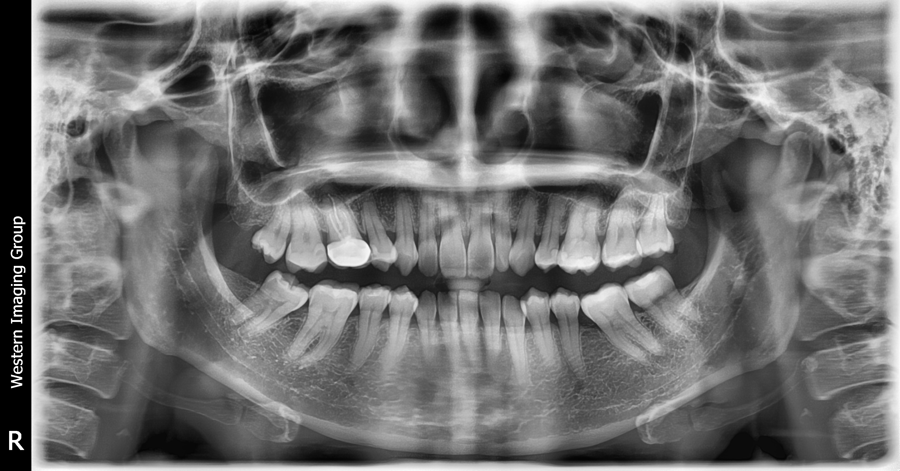 surrounding structures of the mouth e.g. mandible and maxilla.
Why have it?
Your doctor or dentist will let you know why they are referring you for dental imaging; most commonly they are done before having braces fitted or to check for periodontal disease (disease of the tissues).
How long does it take?
Only a few minutes.
What do I have to do?
There is no preparation for this examination, except to remove earrings and necklaces. Instructions will be given to you before commencement of the examination regarding positioning of your head etc.
Where is it performed?
Dental imaging is performed at our Campbell Street and Marsden Park rooms.
If you require any further information, or have any questions, please ask the receptionist, radiographer or radiologist before the examination is performed.
Alternatively, you can contact the practice on 9622 2292 or via email.
What is it?
DXA stands for Dual-energy X-ray Absorptiometry. It takes measurements from the lumbar spine (lower back) and femoral neck (hip) which are then calculated to give the average bone mineral density for both regions.
The scan is able to tell you if your bones show significant demineralisation or if you fit the criteria for osteoporosis.
How long does it take?
Approximately 15 minutes.
What do I have to do?
There is no preparation for this examination.
Where is it performed?
DXA is performed at our Campbell Street rooms.
If you require any further information, or have any questions, please ask the receptionist, radiographer or radiologist before the examination is performed.
Alternatively, you can contact the practice on 9622 2292 or via email.
What is it?
X-rays are a type of electromagnetic radiation that can be used to penetrate the soft tissues of the body. This allows the radiologist to see bone, muscle, fat, fluid and any contrasting material.
 When you think of x-rays it is no doubt general x-rays that you picture.
These include the following:
Chest X-Ray
This looks at the structures of the chest and allows the radiologist to assess the lungs and heart region.
Abdomen X-Ray
This demonstrates the internal structures of the abdomen and allows the radiologist to identify abnormal bowel patterns or assess for kidney stones.
Skull, Sinuses and Facial Bones X-Ray
This allows the radiologist to identify any problems relating to the regions of the head.
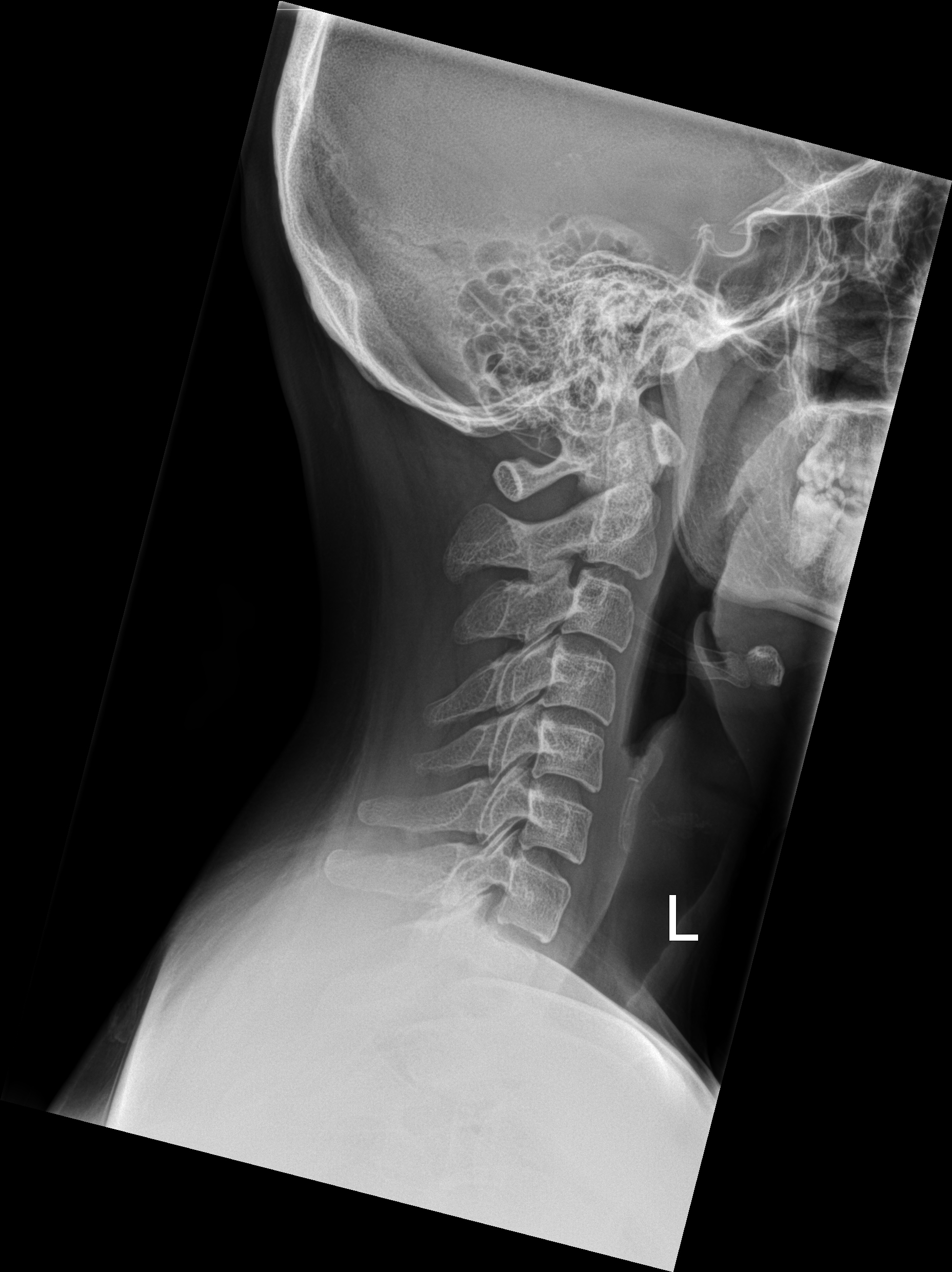
Spinal X-Rays
This includes the cervical spine (neck), thoracic spine (torso) and lumbar spine (lower back) and allows the radiologist to assess the bones of the spine.
Extremities
These include regions such as hands, fingers, toes, feet, knees, ankles etc. and allow the radiologist to assess the bones and soft tissue structures of the area examined.
Where is it performed?
General x-rays are performed at Campbell Street, Marsden Park and Plumpton.
If you require any further information, or have any questions, please ask the receptionist, radiographer or radiologist before the examination is performed.
Alternatively, you can contact the practice on 9622 2292 or via email.
What is it?
A mammogram is a low dose x-ray examination which shows the internal features of your breast. It is one of the  ways to detect breast cancer, often at an early stage before a lump is felt.
Unfortunately not all cancers can be detected by mammography.
Why have it?
The principal reason is to detect breast cancer.
How long does it take?
Between about 15-30 minutes.
What do I have to do?
Before the examination you must ensure you have washed all talcum powder, perfume or deodorant from around the breast area.
Please wear loose fitting clothing – skirt/pants and top are preferable to a dress and the skirt/pants can be left on during the procedure, thus privacy and warmth can be maintained.
What does the procedure involve?
Your breast needs to be compressed as flat as possible to obtain appropriate views of the internal structure. The majority of patients tolerate this procedure very well; however some people can unfortunately find the compression uncomfortable or even painful.
Where is it performed?
Mammograms are performed at our Women's Imaging Centre.
If you require any further information, or have any questions, please ask the receptionist, radiographer or radiologist before the examination is performed.
Alternatively, you can contact the practice on 9622 2292 or via email.
What is it?
An injection of Cortisone can be made (under ultrasound or CT control) into the joints and musculo-skeletal areas.
Why have it?
The injection is for the relief of pain in conditions such as subacromial bursitis (inflammation overlying the shoulder tendons) and plantar fasciitis (inflammation of the plantar fascia connective tissue in the heel).
It can be done in joints such as the shoulder or hip, or into the soft tissue such as the foot. The radiologist will inject a local anaesthetic as well as a steroid (this will sting a little).
How long does it take?
Approximately 15 minutes.
What do I have to do?
There is no preparation for this examination.
Where is it performed?
All MSK and joint injections are performed at our Campbell Street and Marsden Park rooms.
If you require any further information, or have any questions, please ask the receptionist, radiographer or radiologist before the examination is performed.
Alternatively, you can contact the practice on 9622 2292 or via email.
What is it?
Nuchal translucency scanning is a non-invasive prenatal test performed in the first trimester. The ultrasound examination assesses the risk of having a baby with a chromosomal abnormality, such as Trisomy 21, 18 and 13.
 Nuchal translucency scanning must be done early in the pregnancy, between 12 and 14 weeks. The test measures the thickness of a small fluid collection on the back of the baby’s (foetus’) neck. This thickness is measured then analysed by a special computer programme which calculates the risk of having a baby with a chromosomal abnormality.
The remainder of the developing baby (foetus) will also be imaged at the time of the examination.
A blood test is necessary for the results and we will provide you with a referral to have one done.
Nuchal translucency scanning has a sensitivity of about 80% taking into account the mother’s age, other measurements and blood tests. (A low risk result does not mean there is no risk).
Why have it?
The examination is a screening test that provides an estimate value of the likelihood of your baby having a chromsomal defect such as Trisomy 21, 18 or 13.
How long does it take?
The scan itself takes approximately 30 minutes, depending on the position of the baby (foetus). A blood test must then be conducted. Once the blood test results are received, these are collated with the other data received at the time of the scan and full results are produced. All up, results can take up to three working days.
What do I have to do?
There is no preparation required for a Nuchal Translucency ultrasound.
Where is it performed?
Nuchal Translucency ultrasounds are performed at our Campbell Street, Marsden Park and Plumpton rooms.
If you require any further information, or have any questions, please ask the receptionist, radiographer or radiologist before the examination is performed.
Alternatively, you can contact the practice on 9622 2292 or via email.
Dr Matthew Lee specialises in spinal pain therapy and has trained in both Australia as well as in Europe to gain considerable expertise in this area.
Dr Lee is a preferred provider of sacro-iliac joint and back injections for specialists in NSW as well as interstate.
At Western Imaging Group we provide the following services to help manage spinal pain:
If you require any further information, or have any questions, please ask the receptionist, radiographer or radiologist before the examination is performed.
Alternatively, you can contact the practice on 9622 2292 or via email.
What is it?
Ultrasound uses high frequency sound waves to produce a picture/image of your internal organs/anatomy.
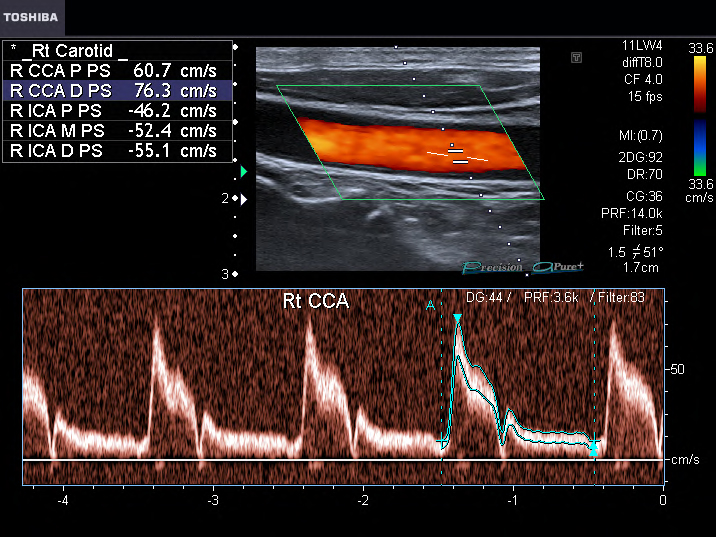 The sound waves (which cannot be heard) produced by the machine bounce off your internal organs and are then detected by the machinery so that an image can be produced on the screen. Doppler ultrasound looks at blood flow (speed and direction) in your arteries and veins. With this form of ultrasound the sound waves can be heard as well as an image being produced.
Why have it?
The reason for the test is best explained to you by your referring doctor. It can be used to diagnose numerous conditions as well as look at the developing baby (foetus).
How long does it take?
It depends on what is being examined – between about 15 minutes to 1 hour.
What do I have to do?
Depending on what part of your body is being examined, there may be no preparation or you may need to fast for up to 6 hours or you may need to drink a number of glasses of water to fill your bladder. The reception staff will explain what is necessary.
Again, depending on what part of your body is being examined, the examination may be carried out standing, sitting or lying. Slippery gel will be put on the part of your body to be examined. This is necessary to help transmit the sound waves in to your body. A transducer/probe will then be put on the gel and the probe will be moved by the sonographer and an image produced.
The examination is not painful, but in some cases you may experience some discomfort from the pressure of the transducer, particularly if it is overlying an area where you are complaining of pain.
Ultrasound examinations performed at Western Imaging Group include general (e.g. pelvic and abdominal ultrasounds), musculoskeletal (e.g. shoulder), doppler (e.g. leg veins or arteries), obstetric (e.g. morphology and dating scans), gynaecological (e.g. female pelvic ultrasound) and small parts (e.g. thyroid).
Where is it done?
Ultrasound is performed at our Campbell Street, Marsden Park and Plumpton rooms.
If you require any further information, or have any questions, please ask the receptionist, radiographer or radiologist before the examination is performed.
Alternatively, you can contact the practice on 9622 2292 or via email.